
 |
Digestion is the mechanical and chemical breakdown of food into smaller components that are more easily absorbed into a blood stream, for instance. |
Anatomy
Because of wide species variations, the digestive system of vertebrates is best described in terms of the headgut, foregut, midgut, pancreas, biliary system, and hindgut. The headgut consists of the mouthparts and pharynx, which serve for the procurement and the initial preparation and swallowing of food. The foregut consists of an esophagus for the swallowing of food and, in most species, a stomach that serves for its storage and initial stages of digestion. The esophagus of most vertebrates is lined with a multilayer of cells that are impermeable to absorption. In most birds it contains the crop, an skyrocketing of its wall that provides for the temporary storage of food. A stomach is present in all but the cyclostomes and some species of advanced fish and in the larval amphibians. In most vertebrates it consists of a dilated segment of the gut that is separated from the esophagus and midgut by muscular sphincters or valves. This is often referred to as a simple stomach. However, in birds these functions are carried out by the crop (storage), proventriculus (secretion), and gizzard (grinding or mastication). In most vertebrates, a major portion of the stomach is lined with a proper gastric mucosa (epithelium), which secretes mucus, hydrochloric acid (HCl), and pepsinogen. The distal (pyloric) part of the stomach secretes mucus and bicarbonate ions (HCO−3), and its muscular contractions help reduce the size of food particles and transfer partially digested food into the midgut. The stomach of reptiles and most mammals has an additional area of cardiac mucosa near its entrance, which also secretes mucus and bicarbonate ions.
The midgut or small intestine is the principal site for the digestion of food and the absorption of nutrients. It is lined with a single layer of cells that secrete mucus and fluids, contain enzymes that aid in the final stages of carbohydrate and protein digestion, and absorb nutrients from the lumen into the circulatory system. The surface area of the lumen can be increased by a variety of means, such as folds and pyloric ceca (blind sacs) in fish. In higher vertebrates the lumen surface is increased by the presence of villi, which are macroscopic projections of the epithelial and subepithelial tissue.
The lumen surface is also expanded by a brush border of microvilli on the lumen-facing (apical) surface of the midgut absorptive cells in all vertebrates. The brush border membranes contain enzymes that aid in the final digestion of food and mechanisms that provide for the selective absorption of nutrients. The luminary surface area of the human small intestine is increased 10-fold by the presence of villi and an additional 20-fold by the microvilli, resulting in a total surface area of 310,000 in.2 (2,000,000 cm2).
Digestion in the midgut is aided by secretions of digestive enzymes and fluid by pancreatic tissue, and secretion of bile by the liver. Pancreatic tissue is distributed along the intestinal wall, and even into the liver, of some species of fish. However, the pancreas is a compact organ in sharks, skates, rays, many teleosts, and all other vertebrates. The liver is a compact organ in all vertebrates. One of its many functions is the secretion of bile. In most vertebrates, the bile is stored in the gallbladder and released into the intestine as needed, but a gallbladder is absent in some species of fish and mammals. Bile salts serve to emulsify lipids and increase their surface area available for digestion by the water-soluble lipase. See also Gallbladder; Liver; Pancreas.
The hindgut is the final site of digestion and absorption prior to defecation or evacuation of waste products. The hindgut of fish, amphibian larvae, and a few mammals is short and difficult to distinguish from the midgut. However, the hindgut of adult amphibians and reptiles, birds, and most mammals is a distinct segment, which is separated from the midgut by a muscular sphincter or valve. It also tends to be larger in diameter. Thus, the midgut and hindgut of these animals are often referred to as the small intestine and the large intestine. See also Intestine.
The hindgut of some reptiles and many mammals includes a blind sac or cecum near its junction with the midgut. A pair of ceca are present in the hindgut of many birds and a few mammalian species. The remainder of the hindgut consists of the colon and a short, straight, terminal segment, which is called the rectum in mammals. The digestive and urinary tracts exit separately from the body of most species of fish and mammals. However, in adult amphibians and the reptiles, birds, and some mammals, this segment terminates in a chamber called the cloaca, which also serves as an exit for the urinary and reproductive systems. The hindgut or, where present, the cloaca terminates in the anus. See also Colon; Urinary system.
The hindgut is similarly lined with a single layer of absorptive and mucus-secreting cells. However, it lacks villi, and (with the exception of the cecum of birds) its absorptive cells lack digestive enzymes and the ability to absorb most nutrients. One major function of the hindgut is to reabsorb the fluids secreted into the upper digestive tract and (in animals that have a cloaca) excreted in the urine. It also serves as the principal site for the microbial production of nutrients in the herbivorous reptiles and birds and in most herbivorous mammals. Thus, the hindgut tends to be longest in animals that need to conserve water in an arid environment, and has a larger capacity in most herbivores.
METHODS OF DIGESTION
The digestive system starts at the mouth and ends at the anus, it is approximately 20 to 30 feet long. Drinking water and eating a healthy diet will keep the digestive system healthy. Almost all animals have a tube-type digestive system in which food enters the mouth, passes through a long tube, and exits as feces (poop) through the anus. The smooth muscle in the walls of the tube-shaped digestive organs rhythmically and efficiently moves the food through the system, where it is broken down into tiny absorber atoms and molecules. During the process of absorption, nutrients that come from the food (including carbohydrates, proteins, fats, vitamins, and minerals) pass through channels in the intestinal wall and into the bloodstream. The blood works to distribute these nutrients to the rest of the body. The waste parts of food that the body can't use are passed out of the body as feces. Every morsel of food we eat has to be broken down into nutrients that can be absorbed by the body, which is why it takes hours to fully digest food. In humans, protein must be broken down into amino acids, starches into simple sugars, and fats into fatty acids and glycerol. The water in our food and drink is also absorbed into the bloodstream to provide the body with the fluid it needs.
  |
The reproductive system or genital system is a system of organs within an organism which work together for the purpose of reproduction.
Asexual Reproduction
Fission, budding, fragmentation, and the formation of rhizomes and stolons are some of the mechanisms that allow organisms to reproduce asexually. The hydra produces buds; starfish can regenerate an entire body from a fragment of the original body. Asexual reproduction allows an organism to rapidly produce many offspring without the time and resources committed to courtship, finding a mate, and mating. The lack of genetic variability in asexually reproducing populations can be detrimental when environmental conditions (for which all the clones are so well adapted) change quickly.
Sexual Reproduction
In sexual reproduction new individuals are produced by the fusion of haploid gametes to form a diploid zygote. Sperm are male gametes, ova (ovum singular) are female gametes. Meiosis produces cells that are genetically distinct from each other; fertilization is the fusion of two such distinctive cells that produces a unique new combination of alleles, thus increasing variation on which natural selection can operate.
Rotifers will reproduce asexually when conditions are favorable by having females produce eggs by mitosis. When conditions deteriorate, rotifers will reproduce sexually and encase their zygotes inside a resistant shell. Once conditions improve, these eggs hatch into diploid individuals. Rotifers thus use sexual reproduction as way to survive a deteriorating environment. Sexual reproduction offers the benefit of generating genetic variation among offspring, which enhances the chances of the population's survival. Costs of this process include the need for two individuals to mate, courtship rituals, as well as a number of basic mechanisms described later. The Male Reproductive System
Testes are suspended outside the abdominal cavity by the scrotum, a pouch of skin that keeps the testes close or far from the body at an optimal temperature for sperm development. Seminiferous tubules are inside each testis, and are where sperm are produced by meiosis. About 250 meters (850 feet) of tubules are packed into each testis. Spermatocytes inside the tubules divide by meiosis to produce spermatids that in turn develop into mature sperm.
 |
Spermatogenesis
Sperm production begins at puberty as it continues throughout life, with several hundred million sperm being produced each day. Once sperm form in which they move into the epididymis, where they mature and are stored.

Male Sex Hormones
The anterior pituitary produces follicle-stimulating hormone (FSH) and luteinizing hormone (LH). Action of LH is controlled by the gonadotropin-releasing hormone (GnRH). LH stimulates cells in the seminiferous tubules to secrete testosterone, which has a role in sperm production and developing male secondary sex characteristics. F SH acts on cells to help in sperm maturation. Negative feedback by testosterone controls the actions of GnRH.

Sexual Structures
Sperm pass through the vas deferens and connect to a short ejaculatory duct that connects to the urethra. The urethra passes through the penis and opens to the outside. Secretions from the seminal vesicles add fructose and prostaglandins to sperm as they pass. The prostate gland secretes a milky alkaline fluid. The bulbourethral gland secretes a mucus-like fluid that provides lubrication for intercourse. Sperm and secretions make up semen.
The Female Reproductive System
The female gonads, ovaries, are located within the lower abdominal cavity.


CIRCULATORY SYSTEM (THE HEART)
The circulatory system is an organ system that passes nutrients (such as amino acids,electrolytes and lymph), gases, hormones, blood cells, etc. to and from cells in the body to help fight diseases, stabilize body temperature and pH, and to maintain homeostasis.
This system may be seen strictly as a blood distribution network, but some consider the circulatory system as composed of the cardiovascular system, which distributes blood, and the lymphatic system, which distributes lymph. While humans, as well as other vertebrates, have a closed cardiovascular system (meaning that the blood never leaves the network of arteries, veins and capillaries), some invertebrate groups have an open cardiovascular system. The most primitive animal phyla lack circulatory systems. The lymphatic system, on the other hand, is an open system.
Two types of fluids move through the circulatory system: blood and lymph. The blood, heart, and blood vessels form the cardiovascular system. The lymph, lymph nodes, and lymph vessels form the lymphatic system. The cardiovascular system and the lymphatic system collectively make up the circulatory system.

The Heart
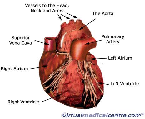
The heart pumps oxygenated blood to the body and deoxygenated blood to the lungs. In the human heart there is one atrium and one ventricle for each circulation, and with both a systemic and a pulmonary circulation there are four chambers in total: left atrium, left ventricle, right atrium and right ventricle. The right atrium is the upper chamber of the right side of the heart. The blood that is returned to the right atrium is deoxygenated (poor in oxygen) and passed into the right ventricle to be pumped through the pulmonary artery to the lungs for re-oxygenation and removal of carbon dioxide. The left atrium receives newly oxygenated blood from the lungs as well as the pulmonary vein which is passed into the strong left ventricle to be pumped through the aorta to the different organs of the body.

Closed cardiovascular system
The cardiovascular systems of humans are closed, meaning that the blood never leaves the network of blood vessels. In contrast, oxygen and nutrients diffuse across the blood vessel layers and enters interstitial fluid, which carries oxygen and nutrients to the target cells, and carbon dioxide and wastes in the opposite direction. The other component of the circulatory system, the lymphatic system, is not closed.
Oxygen transportation
About 98.5% of the oxygen in a sample of arterial blood in a healthy human breathing air at sea-level pressure is chemically combined with haemoglobin molecules. About 1.5% is physically dissolved in the other blood liquids and not connected to haemoglobin. The haemoglobin molecule is the primary transporter of oxygen in mammals and many other species.
 | ||
The heart is cone shaped organ made up of cardiac muscle called myocardium tissue. It is the size of a clenched fist and is responsible for pumping blood around the body. It weighs around 250 to 350 grams.The pericardium is a protective connective tissue enclosing the heart. It is composed of two layers. The outer loosely fitting sac is the fibrous pericardium. It protects and anchors the heart to the surrounding organs. The inner layer is the serous pericardium and it closely adheres to the surface of the heart. Between the outer and inner layer is the pericardial fluid which acts as a lubricant and reduces friction. The heart pumps around 2000 gallons of blood everyday and beats nearly 100,000 times per day.
The human heart is a muscular, cone-shaped, hollow organ about the size of a fist (about 12cm in length and 9cm in breadth). The heart is situated behind the sternum, between the lungs in the thoracic cavity. The heart is tilted slightly such that its apex is towards the left side.
External Heart Anatomy
The major part of the heart is made up of muscles and is called myocardium. The inner lining of the heart is called the endothelium. The heart is covered by a membrane called pericardium. The pericardium encloses the pericardial cavity that houses the heart.
Open circulatory system
The open circulatory system is a system in which fluid (called hemolymph) in a cavity called the hemocoel bathes the organs directly with oxygen and nutrients and there is no distinction between blood and interstitial fluid; this combined fluid is called hemolymph or haemolymph. Muscular movements by the animal during locomotion can facilitate hemolymph movement, but diverting flow from one area to another is limited. When the heart relaxes, blood is drawn back toward the heart through open-ended pores (ostia).
Hemolymph fills all of the interior hemocoel of the body and surrounds all cells. Hemolymph is composed of water, inorganic Salts (mostly Na+, Cl-, K+, Mg2+, and Ca2+), and organic compounds (mostly carbohydrates, proteins, and lipids). The primary oxygen transporter molecule is hemocyanin. There are free-floating cells, the hemocytes, within the hemolymph. They play a role in the arthropod immune system.
Internal View of the Heart
The human heart is four-chambered. The upper chambers are called the atria or the auricles and the lower two chambers are called the ventricles. The two atria are separated by the interatrial septum. The two ventricles are separated from each other by the interventricular septum. The ventricles have more muscular walls than the auricles.
The right side of the heart is concerned with deoxygenated blood and the left side of the heart with the oxygenated blood. The right auricle opens into the lower right ventricle. This opening is guarded by auriculo-ventricular valve (auriculo ventricular valve). This valve is called the tricuspid valve as it has three flaps. The flaps of the valves are connected to the walls of the ventricle by tendons called the chorda tendinae.
Tricuspid Valves
The left auricle opens into the lower left ventricle. This opening is guarded by the bicuspid (having two flaps) or the mitral valve. The tricuspid and the bicuspid valves prevent backflow of blood into the auricles from the ventricles when the latter pump the blood into the blood vessels.
Circulatory systems are absent in some animals, including flatworms (phylum Platyhelminthes). Their body cavity has no lining or enclosed fluid. Instead a muscular pharynx leads to an extensively branched digestive system that facilitates direct diffusion of nutrients to all cells. The flatworm's dorso-ventrally flattened body shape also restricts the distance of any cell from the digestive system or the exterior of the organism. Oxygen can diffuse from the surrounding waterinto the cells, and carbon dioxide can diffuse out. Consequently every cell is able to obtain nutrients, water and oxygen without the need of a transport system.
Some animals, such as jellyfish, have more extensive branching from their gastrovascular cavity (which functions as both a place of digestion and a form of circulation), this branching allows for bodily fluids to reach the outer layers, since the digestion begins in the inner layers.

Bicuspid Valves
The deoxygenated blood from the different parts of the body are collected by the two major veins called the vena cavae - superior vena cava collecting from the upper body and the inferior vena cava collecting from the lower body. The blood from the vena cavae is poured into the right auricle.
The right ventricle opens to a major artery called the pulmonary artery which takes the deoxygenated blood to the lungs. The junction between the pulmonary artery and the right ventricle is guarded by a semilunar valve. This valve prevents the backflow of blood into the ventricle from the artery. The left auricle receives oxygenated blood from the left and right pulmonary veins coming from the left and right lung respectively. The left ventricle opens into a major artery called the aorta. The junction between the aorta and the left ventricle is also guarded by semilunar valve that prevents backflow of blood from the aorta into the ventricle.
The main components of the human cardiovascular system are the heart, blood, and blood vessels.[3] It includes: the pulmonary circulation, a "loop" through the lungs where blood is oxygenated; and the systemic circulation, a "loop" through the rest of the body to provideoxygenated blood. An average adult contains five to six quarts (roughly 4.7 to 5.7 liters) of blood, which consists of plasma, red blood cells, white blood cells, and platelets. Also, the digestive system works with the circulatory system to provide the nutrients the system needs to keep theheart pumping.
Pulmonary circulation
The pulmonary circulatory system is the portion of the cardiovascular system in which oxygen-depleted blood is pumped away from the heart, via the pulmonary artery, to the lungs and returned, oxygenated, to the heart via the pulmonary vein.
Oxygen deprived blood from the vena cava, enters the right atrium of the heart and flows through the tricuspid valve (right atrioventricular valve) into the right ventricle, from which it is then pumped through the pulmonary semilunar valve into the pulmonary artery to the lungs. Gasexchange occurs in the lungs, whereby CO2 is released from the blood, and oxygen is absorbed. The pulmonary vein returns the now oxygen-rich blood to the heart`
Systemic Circulation Systemic circulation is the portion of the cardiovascular system which transports oxygenated blood away from the heart, to the rest of the body, and returns oxygen-depleted blood back to the heart. Systemic circulation is, distance-wise, much longer than pulmonary circulation, transporting blood to every part of the body.
Therefore, the right side of the heart is concerned with the de-oxygenated blood and the left side of the heart is concerned with the oxygenated blood. Further, the auricles are the receiving chambers and the ventricles are the pumping chambers. When the ventricles pump blood into the blood vessels, the bicuspid and tricuspid valves prevent the back flow into the auricles.
Circulatory System of vertebrates
The circulatory systems of all vertebrates, as well as of annelids (for example, earthworms) andcephalopods (squids, octopuses and relatives) are closed, just as in humans. Still, the systems offish, amphibians, reptiles, and birds show various stages of the evolution of the circulatory system.
In fish, the system has only one circuit, with the blood being pumped through the capillaries of thegills and on to the capillaries of the body tissues. This is known as single cycle circulation. The heart of fish is therefore only a single pump (consisting of two chambers).
In amphibians and most reptiles, a double circulatory system is used, but the heart is not always completely separated into two pumps. Amphibians have a three-chambered heart.
In reptiles, the ventricular septum of the heart is incomplete and the pulmonary artery is equipped with a sphincter muscle. This allows a second possible route of blood flow. Instead of blood flowing through the pulmonary artery to the lungs, the sphincter may be contracted to divert this blood flow through the incomplete ventricular septum into the left ventricle and out through the aorta. This means the blood flows from the capillaries to the heart and back to the capillaries instead of to the lungs. This process is useful to ectothermic (cold-blooded) animals in the regulation of their body temperature.
Birds and mammals show complete separation of the heart into two pumps, for a total of four heart chambers; it is thought that the four-chambered heart of birds evolved independently from that of mammals.
Cardiac Cycle
The function of the heart is to pump blood into the blood vessels to ensure that blood reaches all the parts of the body. This is done by the contraction and relaxation of the chambers of the heart. Contraction is called systole and relaxation is called diastole. The pumping action of heart takes place in a rhythmic pattern.
It consists of three stages:
|


Ventilation
In respiratory physiology, ventilation (or ventilation rate) is the rate at which gas enters or leaves the lung. It is categorized under the following definitions:
| Measurement | Equation | Description |
|---|---|---|
| Minute ventilation | tidal volume * respiratory rate[1][2] | the total volume of gas entering the lungs per minute. |
| Alveolar ventilation | (tidal volume - dead space) * respiratory rate [1] | the volume of gas per unit time that reaches the alveoli, the respiratory portions of the lungs where gas exchange occurs. |
| Dead space ventilation | dead space * respiratory rate[3] | the volume of gas per unit time that does not reach these respiratory portions, but instead remains in the airways (trachea, bronchi, etc.). |
Control
Ventilation occurs under the control of the autonomic nervous system from parts of the brain stem, the medulla oblongata and the pons. This area of the brain forms the respiration regulatory center, a series of interconnected brain cells within the lower and middle brain stem which coordinate respiratory movements. The sections are the pneumotaxic center, the apneustic center, and the dorsal and ventral respiratory groups. This section is especially sensitive during infancy, and the neurons can be destroyed if the infant is dropped and/or shaken violently. The result can be death due to "shaken baby syndrome".
Inhalation
Inhalation is initiated by the diaphragm and supported by the external intercostal muscles. Normal resting respirations are 10 to 18 breaths per minute, with a time period of 2 seconds. During vigorous inhalation (at rates exceeding 35 breaths per minute), or in approaching respiratory failure, accessory muscles of respiration are recruited for support. These consist of sternocleidomastoid, platysma, and thescalene muscles of the neck. Pectoral muscles and latissimus dorsi are also accessory muscles.
Under normal conditions, the diaphragm is the primary driver of inhalation. When the diaphragm contracts, the ribcage expands and the contents of the abdomen are moved downward. This results in a larger thoracic volume and negative pressure (with respect to atmospheric pressure) inside the thorax. As the pressure in the chest falls, air moves into the conducting zone. Here, the air is filtered, warmed, and humidified as it flows to the lungs.
During forced inhalation, as when taking a deep breath, the external intercostal muscles and accessory muscles aid in further expanding thethoracic cavity. During inhalation the diaphragm contracts.
Exhalation
Exhalation is generally a passive process; however, active or forced exhalation is achieved by the abdominal and the internal intercostal muscles. During this process air is forced or exhaled out.
The lungs have a natural elasticity: as they recoil from the stretch of inhalation, air flows back out until the pressures in the chest and the atmosphere reach equilibrium.[10]
During forced exhalation, as when blowing out a candle, expiratory muscles including the abdominal muscles and internal intercostal muscles, generate abdominal and thoracic pressure, which forces air out of the lungs.
Gas exchange
The major function of the respiratory system is gas exchange between the external environment and an organism's circulatory system. In humans and other mammals, this exchange facilitates oxygenation of the blood with a concomitant removal of carbon dioxide and other gaseous metabolic wastes from the circulation. As gas exchange occurs, the acid-base balance of the body is maintained as part ofhomeostasis. If proper ventilation is not maintained, two opposing conditions could occur: respiratory acidosis, a life threatening condition, and respiratory alkalosis.
Upon inhalation, gas exchange occurs at the alveoli, the tiny sacs which are the basic functional component of the lungs. The alveolar walls are extremely thin (approx. 0.2 micrometres). These walls are composed of a single layer of epithelial cells (type I and type II epithelial cells) close to the pulmonary capillaries which are composed of a single layer of endothelial cells. The close proximity of these two cell types allows permeability to gases and, hence, gas exchange. This whole mechanism of gas exchange is carried by the simple phenomenon of pressure difference. When the air pressure is high inside the lungs, the air from lungs flow out. When the air pressure is low inside, then air flows into the lungs.
Non-respiratory functions
Lung defense mechanisms
Airway epithelial cells can secrete a variety of molecules that aid in lung defense. Secretory immunoglobulins (IgA), collectins (including Surfactant A and D), defensins and other peptides and proteases, reactive oxygen species, and reactive nitrogen species are all generated by airway epithelial cells. These secretions can act directly as antimicrobials to help keep the airway free of infection. Airway epithelial cells also secrete a variety of chemokines and cytokines that recruit the traditional immune cells and others to site of infections.
Metabolic and endocrine functions of the lungs
In addition to their functions in gas exchange, the lungs have a number of metabolic functions. They manufacture surfactant for local use, as noted above. They also contain a fibrinolytic system that lyses clots in the pulmonary vessels. They release a variety of substances that enter the systemic arterial blood and they remove other substances from the systemic venous blood that reach them via the pulmonary artery.
Prostaglandins are removed from the circulation, but they are also synthesized in the lungs and released into the blood when lung tissue is stretched. The lungs also activate one hormone; the physiologically inactive decapeptide angiotensin I is converted to the pressor, aldosterone-stimulating octapeptide angiotensin II in the pulmonary circulation. The reaction occurs in other tissues as well, but it is particularly prominent in the lungs. Large amounts of the angiotensin-converting enzyme responsible for this activation are located on the surface of the endothelial cells of the pulmonary capillaries. The converting enzyme also inactivates bradykinin.
Circulation time through the pulmonary capillaries is less than 1 s, yet 70% of the angiotensin I reaching the lungs is converted to angiotensin II in a single trip through the capillaries. Four other peptidases have been identified on the surface of the pulmonary endothelial cells.
Vocalization
The movement of gas through the larynx, pharynx and mouth allows humans to speak, or phonate. Vocalization, or singing, in birds occurs via the syrinx, an organ located at the base of the trachea. The vibration of air flowing across the larynx (vocal cords), in humans, and the syrinx, in birds, results in sound. Because of this, gas movement is extremely vital for communication purposes.

Humans and mammals
The respiratory system lies dormant in the human fetus during pregnancy. At birth, the respiratory system becomes fully functional upon exposure to air, although some lung development and growth continues throughout childhood. Pre-term birth can lead to infants with under-developed lungs. These lungs show incomplete development of the alveolar type II cells, cells that produce surfactant.
The lungs of pre-term infants may not function well because the lack of surfactant leads to increased surface tension within the alveoli. Thus, many alveoli collapse such that no gas exchange can occur within some or most regions of an infant's lungs, a condition termed respiratory distress syndrome. Basic scientific experiments, carried out using cells from chicken lungs, support the potential for using steroids as a means of furthering development of type II alveolar cells.[11] In fact, once a pre-mature birth is threatened, every effort is made to delay the birth, and a series ofsteroid shots is frequently administered to the mother during this delay in an effort to promote lung growth.
Disorders of the respiratory system can be classified into four general areas:
- Obstructive conditions (e.g., emphysema, bronchitis, asthma)
- Restrictive conditions (e.g., fibrosis, sarcoidosis, alveolar damage, pleural effusion)
- Vascular diseases (e.g., pulmonary edema, pulmonary embolism, pulmonary hypertension)
- Infectious, environmental and other "diseases" (e.g., pneumonia, tuberculosis, asbestosis, particulate pollutants):
Coughing is of major importance, as it is the body's main method to remove dust, mucus, saliva, and other debris from the lungs. Inability to cough can lead to infection. Deep breathing exercises may help keep finer structures of the lungs clear from particulate matter, etc.
The respiratory tract is constantly exposed to microbes due to the extensive surface area, which is why the respiratory system includes many mechanisms to defend itself and prevent pathogens from entering the body.
Disorders of the respiratory system are usually treated internally by a pulmonologist and Respiratory Therapist.
MUSCULAR SYSTEM
The muscular system is an organ system consisting of skeletal, smooth and cardiacmuscles. It permits movement of the body, maintains posture, and circulates blood throughout the body. The muscular system in vertebrates is controlled through the nervous system, although some muscles (such as the cardiac muscle) can be completely autonomous.

Muscles
There are three distinct types of muscles: skeletal muscles, cardiac or heart muscles, and smooth (non-striated) muscles. Muscles provide strength, balance, posture, movement and heat for the body to keep warm.
Upon stimulation by an action potential, skeletal muscles perform a coordinated contraction by shortening each sarcomere. The best proposed model for understanding contraction is the sliding filament model of muscle contraction. Actin and myosin fibers overlap in a contractile motion towards each other. Myosin filaments have club-shaped heads that project toward the actin filaments.
Larger structures along the myosin filament called myosin heads are used to provide attachment points on binding sites for the actin filaments. The myosin heads move in a coordinated style, they swivel toward the center of the sarcomere, detach and then reattach to the nearest active site of the actin filament. This is called a rachet type drive system. This process consumes large amounts of adenosine triphosphate (ATP).
Energy for this comes from ATP, the energy source of the cell. ATP binds to the cross bridges between myosin heads and actin filaments. The release of energy powers the swiveling of the myosin head. Muscles store little ATP and so must continuously recycle the discharged adenosine diphosphate molecule (ADP) into ATP rapidly. Muscle tissue also contains a stored supply of a fast acting recharge chemical, creatine phosphate which can assist initially producing the rapid regeneration of ADP into ATP.
Calcium ions are required for each cycle of the sarcomere. Calcium is released from the sarcoplasmic reticulum into the sarcomere when a muscle is stimulated to contract. This calcium uncovers the actin binding sites. When the muscle no longer needs to contract, the calcium ions are pumped from the sarcomere and back into storage in the sarcoplasmic reticulum.

Anatomy
There are approximately 639 skeletal muscles in the human body.
The following are some major muscles and their basic features:

Aerobic and Anaerobic Muscle Activity
At rest, the body produces the majority of its ATP aerobically in the mitochondria without producing lactic acid or other fatiguing byproducts. During exercise, the method of ATP production varies depending on the fitness of the individual as well as the duration, and intensity of exercise. At lower activity levels, when exercise continues for a long duration (several minutes or longer), energy is produced aerobically by combining oxygen with carbohydrates and fats stored in the body.
Activity that is higher in intensity, with possible duration decreasing as intensity increases, ATP production can switch to anaerobic pathways, such as the use of the creatine phosphate and the phosphagen system or anaerobic glycolysis. Aerobic ATP production is biochemically much slower and can only be used for long-duration, low intensity exercise, but produces no fatiguing waste products that can not be removed immediately from sarcomere and body and results in a much greater number of ATP molecules per fat or carbohydrate molecule. Aerobic training allows the oxygen delivery system to be more efficient, allowing aerobic metabolism to begin quicker. Anaerobic ATP production produces ATP much faster and allows near-maximal intensity exercise, but also produces significant amounts of lactic acid which render high intensity exercise unsustainable for greater than several minutes. The phosphagen system is also anaerobic, allows for the highest levels of exercise intensity, but intramuscular stores of phosphocreatine are very limited and can only provide energy for exercises lasting up to ten seconds. Recovery is very quick, with full creatine stores regenerated within five minutes.

Cardiac Muscle(Heart Muscle)
Heart muscles are distinct from skeletal muscles because the muscle fibers are laterally connected to each other. Furthermore, just as with smooth muscles, they are not controlling themselves. Heart muscles are controlled by the sinus node influenced by the autonomic nervous system.

Smooth Muscle
Smooth muscles are controlled directly by the autonomic nervous system and are involuntary, meaning that they are incapable of being moved by conscious thought. Functions such as heart beat and lungs (which are capable of being willingly controlled, be it to a limited extent) are involuntary muscles but are not smooth muscles.

Control Of Muscle Contraction
Neuromuscular junctions are the focal point where a motor neuron attaches to a muscle. Acetylcholine, (a neurotransmitter used in skeletal muscle contraction) is released from the axon terminal of the nerve cell when an action potential reaches the microscopic junction, called asynapse. A group of chemical messengers cross the synapse and stimulate the formation of electrical changes, which are produced in the muscle cell when the acetylcholine binds to receptors on its surface. Calcium is released from its storage area in the cell's sarcoplasmic reticulum. An impulse from a nerve cell causes calcium release and brings about a single, short muscle contraction called a muscle twitch. If there is a problem at the neuromuscular junction, a very prolonged contraction may occur, tetanus. Also, a loss of function at the junction can produce paralysis.

Skeletal Muscles
Skeletal muscles are organized into hundreds of motor units, each of which involves a motor neuron, attached by a series of thin finger-like structures called axon terminals.
These attach to and control discrete bundles of muscle fibers. A coordinated and fine tuned response to a specific circumstance will involve controlling the precise number of motor units used. While individual muscle units contract as a unit, the entire muscle can contract on a predetermined basis due to the structure of the motor unit. Motor unit coordination, balance, and control frequently come under the direction of the cerebellum of the brain. This allows for complex muscular coordination with little conscious effort, such as when one drives a car without thinking about the process.
These attach to and control discrete bundles of muscle fibers. A coordinated and fine tuned response to a specific circumstance will involve controlling the precise number of motor units used. While individual muscle units contract as a unit, the entire muscle can contract on a predetermined basis due to the structure of the motor unit. Motor unit coordination, balance, and control frequently come under the direction of the cerebellum of the brain. This allows for complex muscular coordination with little conscious effort, such as when one drives a car without thinking about the process.
THE HUMAN BREAST
The breast is the upper ventral region of the torso of a primate, in left and right sides, which in a female contains the mammary gland that secretes milk used to feed infants.
Both men and women develop breasts from the same embryological tissues. However, atpuberty, female sex hormones, mainly estrogen, promote breast development, which does not occur in men, due to the higher amount of testosterone. As a result, women's breasts become far more prominent than those of men.
 |
Morphology
The human breast has two aspects — the functional and the anatomic aspect.
The functional breast
The breast is an apocrine gland that produces milk to feed an infant child; for which the nipple of the breast is centred in (surrounded by) an areola (nipple-areola complex, NAC), the skin color of which varies from pink to dark brown, and has many sebaceous glands. The basic units of the breast are the terminal duct lobular units (TDLUs), which produce the fatty breast milk. They give the breast its offspring-feeding functions as a mammary gland. They are distributed throughout the body of the breast; approximately two-thirds of the lactiferous tissue is within 30-mm of the base of the nipple. The terminal lactiferous ducts drain the milk from TDLUs into 4–18 lactiferous ducts, which drain to the nipple; the milk-glands-to-fat ratio is 2:1 in alactating woman, and 1:1 in a non-lactating woman. In addition to the milk glands, the breast also is composed of connective tissues (collagen, elastin), white fat, and the suspensory Cooper's ligaments. Sensation in the breast is provided by theperipheral nervous system innervation, by means of the front (anterior) and side (lateral) cutaneous branches of the fourth-, the fifth-, and the sixth intercostal nerves, while the T-4 nerve (Thoracic spinal nerve 4), which innervates the dermatomic area, supplies sensation to the nipple-areola complex.
The anatomic breast
A woman’s breasts overlay the pectoralis major muscles and usually extend from the level of the second rib to the level of the sixth rib in the front of the human rib cage; thus, the breasts cover much of the chest area and the chest walls. At the front of the chest, the breast tissue can extend from the clavicle (collarbone) to the middle of the sternum (breastbone). At the sides of the chest, the breast tissue can extend into the axilla (armpit), and can reach as far to the back as the latissimus dorsi muscle, extending from the lower back to thehumerus bone (the longest bone of the upper arm). As a mammary gland, the breast is an inhomogeneous anatomic structure composed of layers of different types of tissue, among which predominate two types: (i) adipose tissue and (ii) glandular tissue, which effects the lactationfunctions of the breasts.
Morphologically, the breast is a cone with the base at the chest wall, and the apex at the nipple, the center of the NAC (nipple-areola complex). The superficial tissue layer (superficial fascia) is separated from the skin by 0.5–2.5 cm of subcutaneous fat (adipose tissue). The suspensory Cooper’s ligaments are fibrous-tissue prolongations that radiate from the superficial fascia to the skin envelope. The adult breast contains 14–18 irregular lactiferous lobes that converge to the nipple, to ducts 2.0–4.5 mm in diameter; the milk ducts (lactiferous ducts) are immediately surrounded with dense connective tissue that functions as a support framework. The glandular tissue of the breast is biochemically supported with estrogen; thus, when a woman reaches menopause (cessation of menstruation) and her body estrogen levels decrease, the milk gland tissue then atrophies, withers, and disappears, resulting in a breast composed of adipose tissue, superficial fascia, suspensory ligaments, and the skin envelope.
The dimensions and the weight of the breast vary among women, ranging approximately 500–1,000 gm (1.1-2.2 pounds) each; thus, a small-to-medium-sized breast weighs 500 gm (1.1 pounds) or less; and a large breast weighs approximately 750–1,000 gm (1.7-2.2 pounds.) The tissue composition ratios of the breast likewise vary among women; some breasts have greater proportions of glandular tissue than of adipose or connective tissues, and vice versa; therefore the fat-to-connective-tissue ratio determines the density (firmness) of the breast. In the course of a woman’s life, her breasts will change size, shape, and weight, because of the hormonal bodily changes occurred in thelarche (pubertal breast development), menstruation (fertility), pregnancy (reproduction), the breast-feeding of an infant child, and the climacterium (the end of fertility).
|
 |
Lymphatic drainage
Approximately 75 per cent of the lymph from the breast travels to the ipsilateral (same-side) axillary lymph nodes, whilst 25 per cent of the lymph travels to the parasternal nodes (beside the sternum bone), to the other breast, and to the abdominal lymph nodes. The axillary lymph nodes include the pectoral (chest), subscapular (under the scapula), and humeral (humerus-bone area) lymph-node groups, which drain to the central axillary lymph nodes and to the apical axillary lymph nodes. The lymphatic drainage of the breasts is especially relevant to oncology, because breast cancer is a cancer common to the mammary gland, and cancer cells can metastasize (break away) from a tumour and be dispersed to other parts of the woman’s body by means of the lymphatic system.
Shape and Support
The morphologic variations in the size, shape, volume, tissue density, pectoral locale, and spacing of the breasts determine their natural shape, appearance, and configuration upon the chest of a woman; yet such features do not indicate its mammary-gland composition (fat-to-milk-gland ratio), nor the potential for nursing an infant child. The size and the shape of the breasts are influenced by normal-life hormonal changes (thelarche, menstruation,pregnancy, menopause) and medical conditions (e.g. virginal breast hypertrophy).[11] The shape of the breasts is naturally determined by the support of the suspensory Cooper's ligaments, the underlying muscle and bone structures of the chest, and the skin envelope. The supensory ligaments sustain the breast from the clavicle (collarbone) and the clavico-pectoral fascia (collarbone and chest), by traversing and encompassing the fat and milk-gland tissues, the breast is positioned, affixed to, and supported upon the chest wall, while its shape is established and maintained by the skin envelope.
The base of each breast is attached to the chest by the deep fascia over the pectoralis major muscles. The space between the breast and the pectoralis major muscle is called retromammary space and gives mobility to the breast. Some breasts are mounted high upon the chest wall, are of rounded shape, and project almost horizontally from the chest, which features are common to girls and women in the early stages of thelarchic development, the sprouting of the breasts. In the high-breast configuration, the dome-shaped and the cone-shaped breast is affixed to the chest at the base, and the weight is evenly distributed over the base area. In the low-breast configuration, a proportion of the breast weight is supported by the chest, against which rests the lower surface of the breast, thus is formed the inframammary fold (IMF). Because the base is deeply affixed to the chest, the weight of the breast is distributed over a greater area, and so reduces the weight-bearing strain upon the chest, shoulder, and back muscles that bear the weight of the bust.
The chest (thoracic cavity) progressively slopes outwards from the thoracic inlet (atop the breastbone) and above to the lowest ribs that support the breasts. The inframammary fold, where the lower portion of the breast meets the chest, is an anatomic feature created by the adherence of the breast skin and the underlying connective tissues of the chest; the IMF is the lower-most extent of the anatomic breast. In the course of thelarche, some girls develop breasts the lower skin-envelope of which touches the chest below the IMF, and some girls do not; both breast anatomies are statistically normal morphologic variations of the size and shape of women's breasts.
|
 | ||||||||||||
Development
The basic morphological structure of the human breast — female and male — is determined during theprenatal development stage. For a girl in puberty, during thelarche (the breast-development stage), the female sex hormones (principally estrogens) promote the sprouting, growth, and development of the breasts, in the course of which, as mammary glands, they grow in size and volume, and usually rest on her chest; these development stages of secondary sex characteristics (breasts, pubic hair, etc.) are illustrated in the five-stage Tanner Scale. During thelarche, the developing breasts sometimes are of unequal size, and usually the left breast is slightly larger; said condition of asymmetry is transitory and statistically normal to female physical and sexual development. Moreover, breast development sometimes is abnormal, manifested either as overdevelopment (e.g. virginal breast hypertrophy) or as underdevelopment (e.g. tuberous breast deformity) in girls and women; and manifested in boys and men as gynecomastia(woman's breasts), the consequence of a biochemical imbalance between the normal levels of the estrogenand testosterone hormones of the male body.
Asymmetry
Approximately two years after the pubertal occurrence of a girl’s first menstrual cycle, the hormoneoestrogen stimulates the development and growth of the glandular, fat, and suspensory tissues that compose the breast; and continues for approximately four years until establishing the final shape of the breast (size, volume, density) when she is a woman of approximately 21 years of age.
Approximately 90 per cent of women possess breasts that are asymmetrical, either in size, volume, or relative position upon the chest; whilst notable breast asymmetry — a breast-size difference of at least one brassière-cup size — is present in 25 per cent of women.
For approximately 5–10% of women, the asymmetry of the breasts is notably different, with the left breast bigger in 62% of the women. The asymmetry can be manifested in the size of the breast, the position of the nipple-areola complex (NAC), the angle of the breast, and the position of the inframammary fold, where the breast meets the chest. The usual resolution for such conditions is corrective mammoplasty, such as mastopexy, breast reduction, or breast augmentation, which treatment is determined by the particular form of breast asymmetry to correct.
IMMUNE SYSTEMS
An immune system is a system of biological structures and processes within an organismthat protects against disease. In order to function properly, an immune system must detect a wide variety of agents, from viruses to parasitic worms, and distinguish them from the organism's own healthy tissue.
Pathogens can rapidly evolve and adapt to avoid detection and destruction by the immune system. As a result, multiple defense mechanisms have also evolved to recognize and neutralize pathogens. Even simple unicellular organisms such as bacteria possess a rudimentary immune system, in the form of enzymes which protect against bacteriophage infections. Other basic immune mechanisms evolved in ancient eukaryotes and remain in their modern descendants, such as plants and insects. These mechanisms include phagocytosis, antimicrobial peptides called defensins, and the complement system. Jawed vertebrates, including humans, have even more sophisticated defense mechanisms, including the ability to adapt over time to recognize specific pathogens more efficiently. Adaptive (or acquired) immunity creates immunological memory after an initial response to a specific pathogen, leading to an enhanced response to subsequent encounters with that same pathogen. This process of acquired immunity is the basis of vaccination.
Disorders of the immune system can result in autoimmune diseases, inflammatory diseasesand cancer. Immunodeficiency occurs when the immune system is less active than normal, resulting in recurring and life-threatening infections. In humans, immunodeficiency can either be the result of a genetic disease, such as severe combined immunodeficiency, or acquired conditions such as HIV/AIDS or the use of immunosuppressive medication. In contrast,autoimmunity results from an hyperactive immune system attacking normal tissues as if they were foreign organisms. Common autoimmune diseases include Hashimoto's thyroiditis, rheumatoid arthritis, diabetes mellitus type 1, and systemic lupus erythematosus. Immunologycovers the study of all aspects of the immune system.

History Of Immunology
Immunology is a science that examines the structure and function of the immune system. It originates from medicine and early studies on the causes of immunity to disease. The earliest known reference to immunity was during the plague of Athens in 430 BC. Thucydides noted that people who had recovered from a previous bout of the disease could nurse the sick without contracting the illness a second time. In the 18th century, Pierre-Louis Moreau de Maupertuis made experiments with scorpion venom and observed that certain dogs and mice were immune to this venom. This and other observations of acquired immunity were later exploited by Louis Pasteur in his development of vaccination and his proposed germ theory of disease. Pasteur's theory was in direct opposition to contemporary theories of disease, such as the miasma theory. It was not until Robert Koch's 1891 proofs, for which he was awarded a Nobel Prize in 1905, that microorganisms were confirmed as the cause of infectious disease. Viruses were confirmed as human pathogens in 1901, with the discovery of the yellow fever virus byWalter Reed.
Immunology made a great advance towards the end of the 19th century, through rapid developments, in the study of humoral immunity and cellular immunity. Particularly important was the work of Paul Ehrlich, who proposed the side-chain theory to explain the specificity of the antigen-antibody reaction; his contributions to the understanding of humoral immunity were recognized by the award of a Nobel Prize in 1908, which was jointly awarded to the founder of cellular immunology, Elie Metchnikoff.
Layered Defense
The immune system protects organisms from infection with layered defenses of increasing specificity. In simple terms, physical barriers prevent pathogens such as bacteria and viruses from entering the organism. If a pathogen breaches these barriers, the innate immune system provides an immediate, but non-specific response. Innate immune systems are found in all plants and animals.[11] If pathogens successfully evade the innate response, vertebrates possess a second layer of protection, the adaptive immune system, which is activated by the innate response. Here, the immune system adapts its response during an infection to improve its recognition of the pathogen. This improved response is then retained after the pathogen has been eliminated, in the form of an immunological memory, and allows the adaptive immune system to mount faster and stronger attacks each time this pathogen is encountered.[12]
Both innate and adaptive immunity depend on the ability of the immune system to distinguish between self and non-self molecules. Inimmunology, self molecules are those components of an organism's body that can be distinguished from foreign substances by the immune system.[13] Conversely, non-self molecules are those recognized as foreign molecules. One class of non-self molecules are called antigens(short for antibody generators) and are defined as substances that bind to specific immune receptors and elicit an immune response.
Surface Barriers
Several barriers protect organisms from infection, including mechanical, chemical, and biological barriers. The waxy cuticle of many leaves, the exoskeleton of insects, the shells and membranes of externally deposited eggs, and skin are examples of mechanical barriers that are the first line of defense against infection.[14] However, as organisms cannot be completely sealed against their environments, other systems act to protect body openings such as the lungs, intestines, and the genitourinary tract. In the lungs, coughing and sneezing mechanically eject pathogens and other irritants from the respiratory tract. The flushing action of tears and urine also mechanically expels pathogens, whilemucus secreted by the respiratory and gastrointestinal tract serves to trap and entangle microorganisms.[15]
Chemical barriers also protect against infection. The skin and respiratory tract secrete antimicrobial peptides such as the β-defensins.[16]Enzymes such as lysozyme and phospholipase A2 in saliva, tears, and breast milk are also antibacterials.[17][18] Vaginal secretions serve as a chemical barrier following menarche, when they become slightly acidic, while semen contains defensins and zinc to kill pathogens.[19][20] In the stomach, gastric acid and proteases serve as powerful chemical defenses against ingested pathogens.
Within the genitourinary and gastrointestinal tracts, commensal flora serve as biological barriers by competing with pathogenic bacteria for food and space and, in some cases, by changing the conditions in their environment, such as pH or available iron.[21] This reduces the probability that pathogens will be able to reach sufficient numbers to cause illness. However, since most antibiotics non-specifically target bacteria and do not affect fungi, oral antibiotics can lead to an "overgrowth" of fungi and cause conditions such as a vaginal candidiasis (a yeast infection).[22] There is good evidence that re-introduction of probiotic flora, such as pure cultures of the lactobacilli normally found in unpasteurized yogurt, helps restore a healthy balance of microbial populations in intestinal infections in children and encouraging preliminary data in studies on bacterial gastroenteritis, inflammatory bowel diseases, urinary tract infection and post-surgical infections.[23][24][25]
|

Hey friends,families and people of the world, I did this mainly to help me and you know somethings that science especially ANATOMY can do. Anatomy is really fun and I hope all of you will agree with me. Even if you don't agree, remember that "I can make it but cannot do it all so lets come together as one and share our knowledge about science around the globe. THANK YOU.
ReplyDelete